Jinchun Wu, PhD
Genome rearrangements have been widely observed in human cancers. Recent whole-genome sequencing data has identified chromothripsis, an event that introduces massive genome rearrangements in only one or a few chromosomes through catastrophic shattering and random reattachment, as one of the most frequent genome rearrangements. Chromothripsis has been associated with poor clinical outcomes in multiple cancers, but the shattering mechanisms that induce chromosome fragmentation remain uncharacterized. Dr. Wu [Marion Abbe Fellow] aims to determine the role of cytoplasmic nucleases (enzymes that cleave DNA) in chromosome shattering and genome rearrangement, which will contribute to our understanding of chromothripsis in all cancers. She will extend this project to a mouse model of glioma to determine the effects of candidate nucleases on cancer progression. Dr. Wu received her PhD and BS from Peking University, Beijing.
Simon Sretenovic, PhD
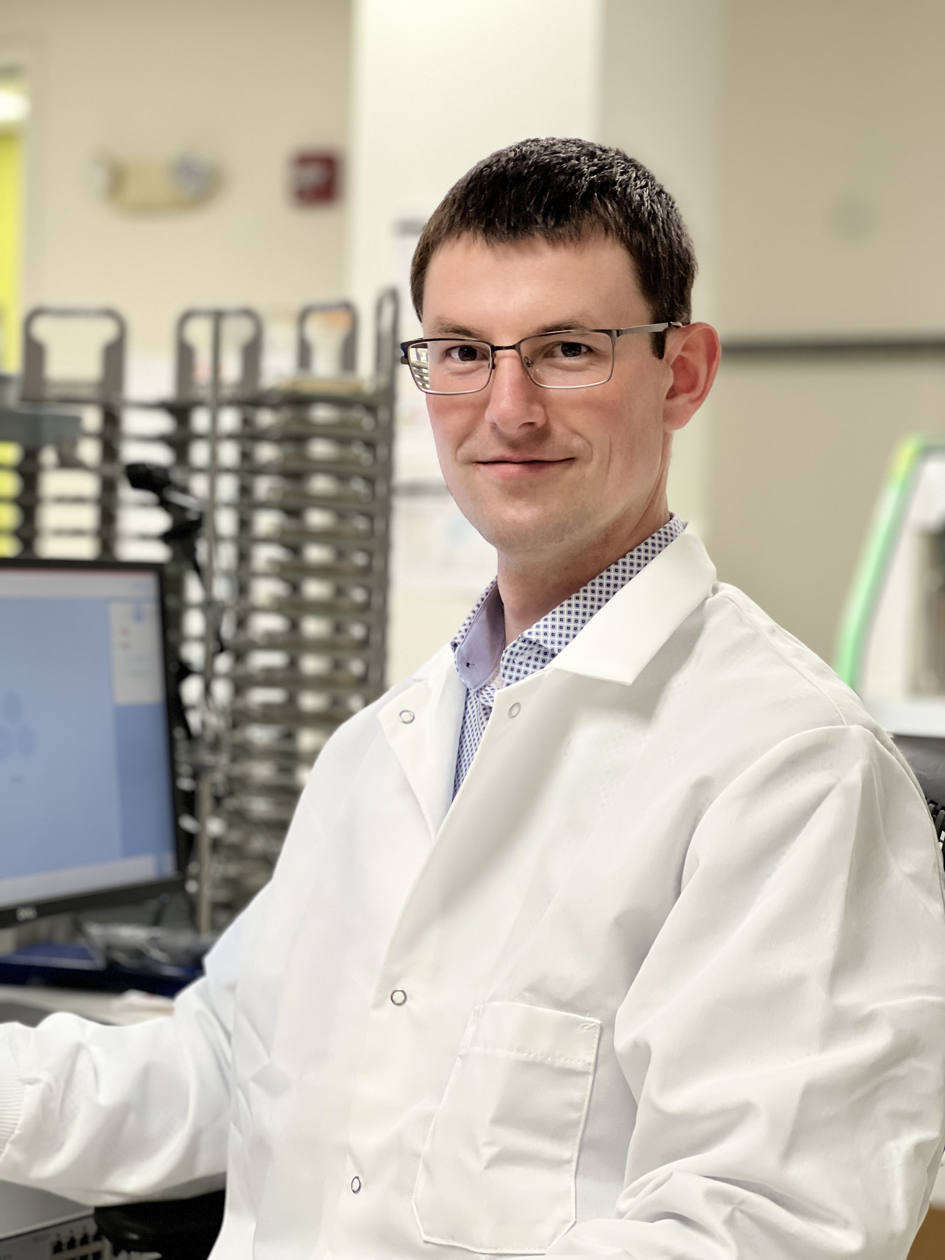
More than one third of all people will receive a cancer diagnosis at some point in their lifetime. Dr. Sretenovic [Connie and Bob Lurie Fellow] is using both yeast and human cell lines to model various properties of cancerous cells as complex genetic traits. Combining novel CRISPR genome editing approaches with next-generation sequencing technology, he aims to dissect the intricate relationships between genetic variants, chemical and physical environmental factors, and phenotypic outcomes (i.e., observable characteristics). The goal of his project is to understand the genetic basis for a panel of cancer-related traits to inform the development of anti-cancer treatments. Dr. Sretenovic received his PhD from the University of Maryland, College Park, and his MS and BS from University of Ljubljana, Ljubljana.
Wenzhi Song, PhD
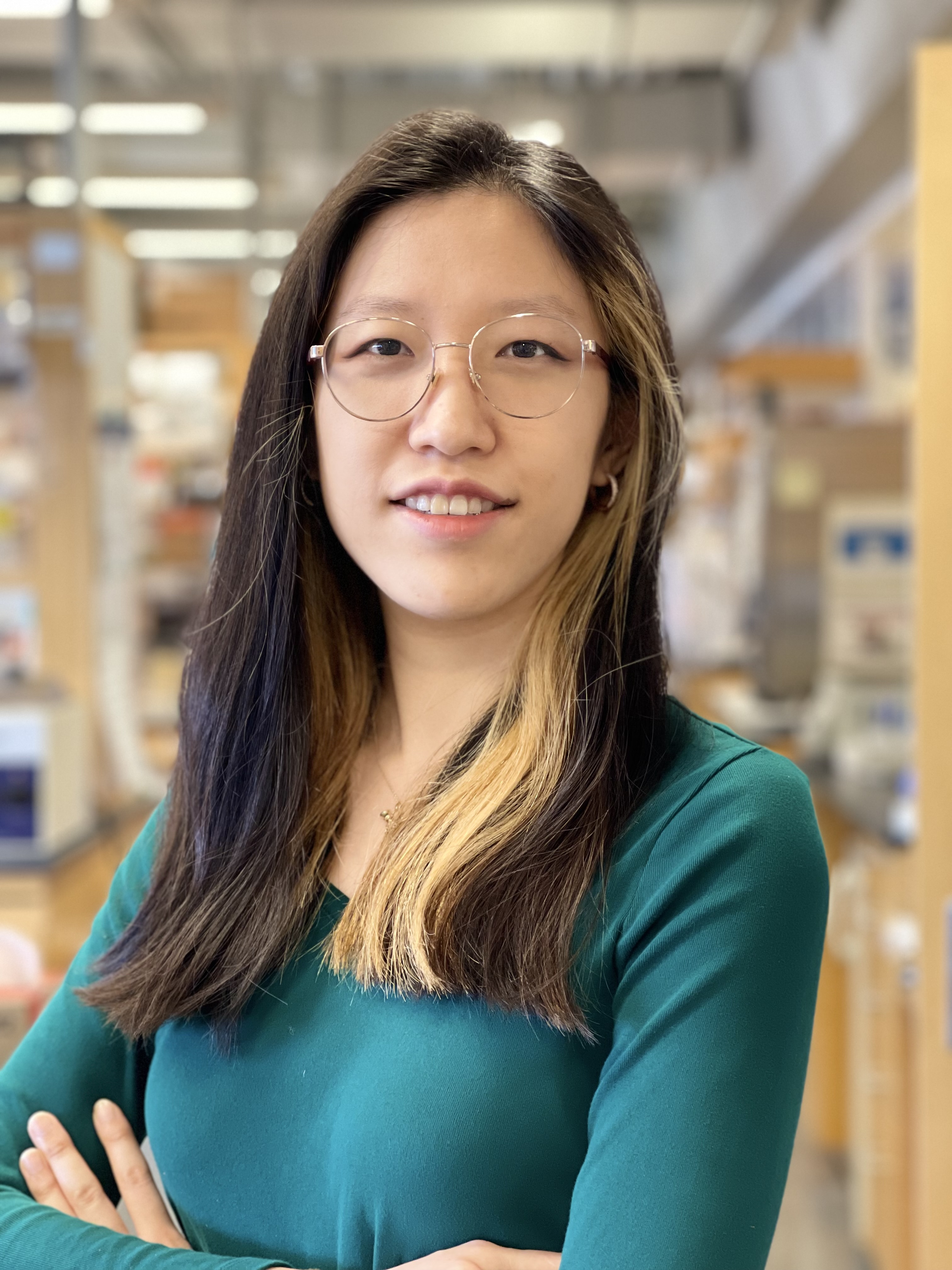
The interaction between cancer cells and their non-malignant neighbors in the tumor microenvironment is critical for cancer progression. While certain types of cellular crosstalk within the tissue safeguard against malignancy, cancer cells are often able to exploit nearby cells to fuel tumor growth. Dr. Song [HHMI Fellow] is interested in understanding how the complex cellular communication network in the skin, namely its sensory and immunological components, contributes to the development of cutaneous squamous cell carcinoma, one of the most common skin cancers. Identifying novel neuronal and immunological interactions within the tumor microenvironment has the potential to uncover pathways regulating cancer progression and anti-tumor immunity. Dr. Song received her PhD from Yale University, New Haven and her AB from Bryn Mawr College, Bryn Mawr.
Yoshiki Sakai, PhD
Normally, epithelial tissues, which cover all external body surfaces and line internal cavities, expel unwanted cells to maintain health in a process known as cell extrusion. However, some cancer cells, particularly those with the common RasV12 mutation, manage to avoid extrusion. Using Drosophila (fruit flies) as a model, Dr. Sakai [Rhee Family Fellow] will explore how RasV12-mutant cells manipulate neighboring cells to avoid extrusion. Understanding this process could lead to new ways to prevent cancer cells from escaping the epithelial defense, offering potential new treatments. Dr. Sakai received his PhD and BS from Nagoya University, Nagoya.
Ian J. Roney, PhD
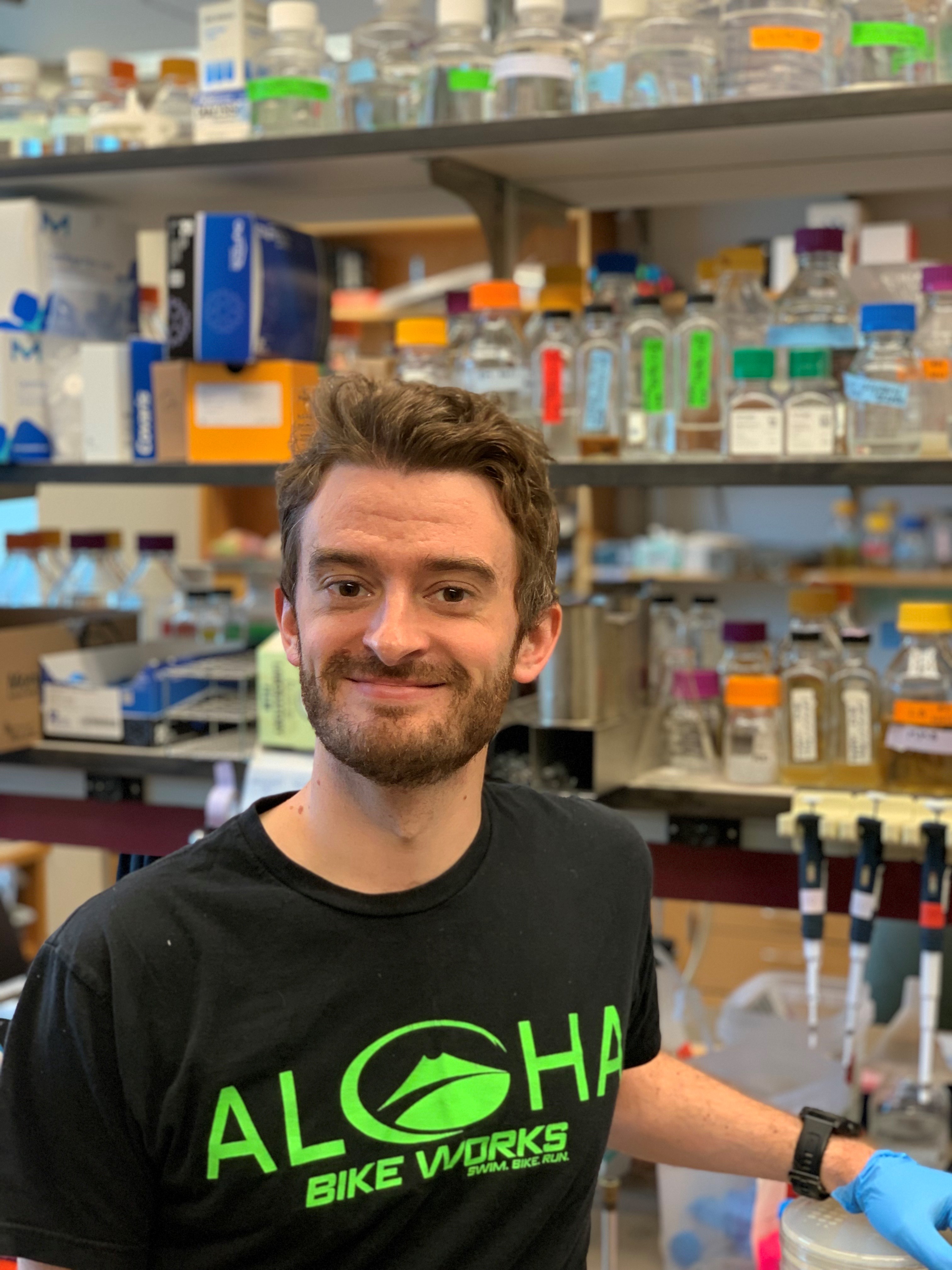
Bacteria have diverse immune systems to defend themselves against viral invaders, many of which use molecular mechanisms also seen in mammalian immune systems. Dr. Roney [HHMI Fellow] studies how bacterial immune systems detect virally compromised cells, and how viruses undermine immune systems to prevent the elimination of virally compromised cells from the population. The goal of his research is to uncover novel mechanisms and principles of immune systems that are found across domains of life. The discoveries resulting from this work will broaden our understanding of how immune systems detect and eliminate compromised cells, like cancer cells, and could help guide development of new immunotherapies. Dr. Roney received his PhD from Harvard University, Cambridge and his MS and BS from University of Ottawa, Ottawa.
David S. Roberts, PhD
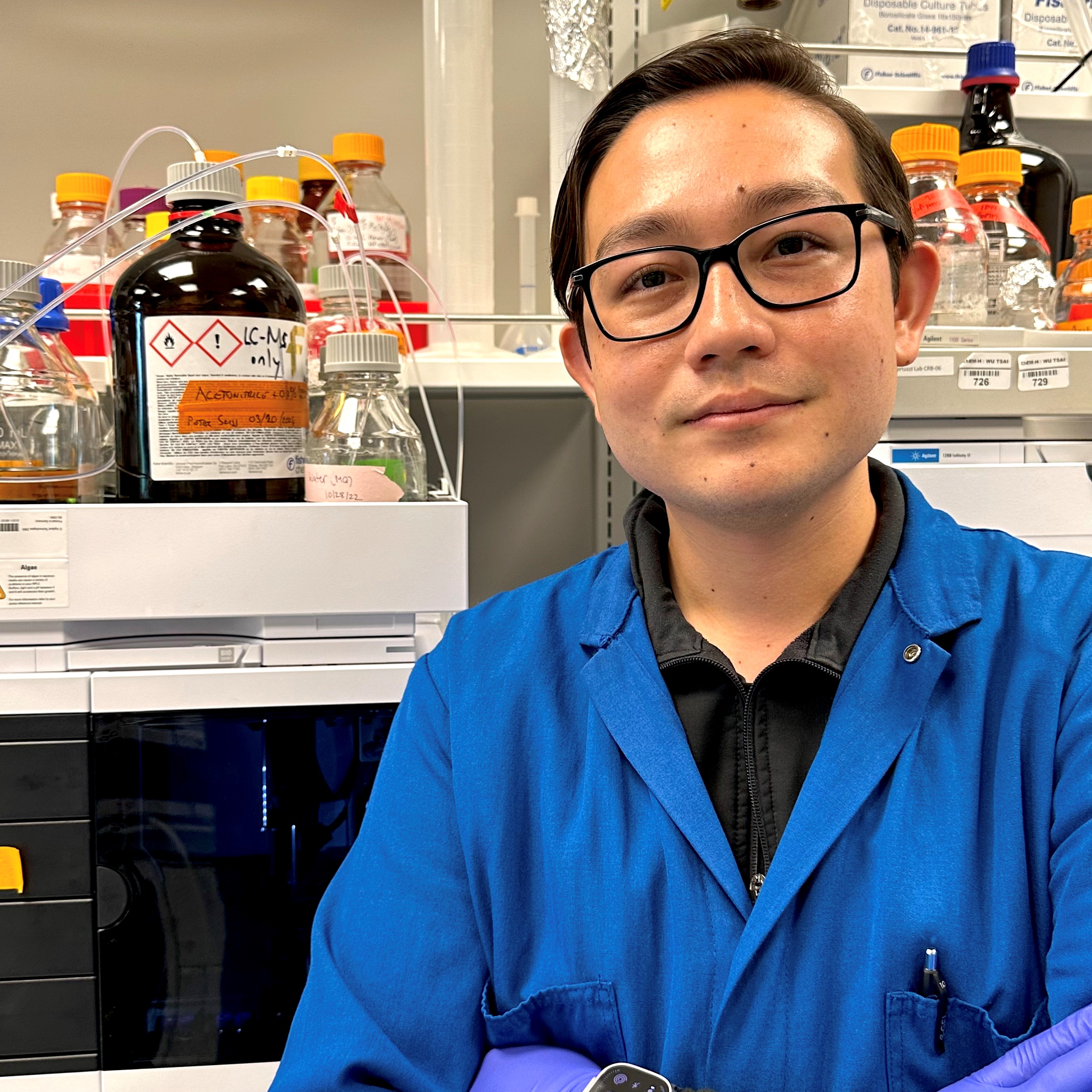
Cancer immunotherapies have shown remarkable benefits, but many tumors remain unresponsive to existing treatments. The mechanisms cancer cells use to evade immune responses during treatment remain largely unknown. Altered cell surface glycosylation, the process of attaching sugars to cell surface biomolecules, is a hallmark of many human cancers. The interaction between cell surface glycoproteins on immune cells with cancer cells represents a major axis of immune evasion and plays a vital role in how cancer cells suppress immune responses during cancer treatment. Dr. Roberts’ [Connie and Bob Lurie Fellow] research aims to molecularly define cell surface glycosylation and understand the role of glycosylation in driving cancer immunosuppression. This knowledge will be leveraged to illuminate the underlying mechanisms of tumor immune evasion and enable next-generation classes of cancer immunotherapies. Dr. Roberts received his PhD from University of Wisconsin–Madison, Madison and his BS from University of California, San Diego.
Expery O. Omollo, PhD

Dr. Omollo [Robert A. Swanson Family Fellow] studies how bacteria have evolved to achieve precise gene expression using strategically placed transcription terminators. In cancer cells, specific mutations lead to uncontrolled transcription of certain genes, resulting in elevated gene expression that fuels cancer progression. Using bacteria as a model, Dr. Omollo aims to uncover how RNA polymerases in cancer cells evade termination signals to maintain high levels of gene expression, encouraging cancer spread. Dr. Omollo received his PhD from University of Wisconsin-Madison, Madison and his BS from Michigan State University, Lansing.
Chaiheon Lee, PhD
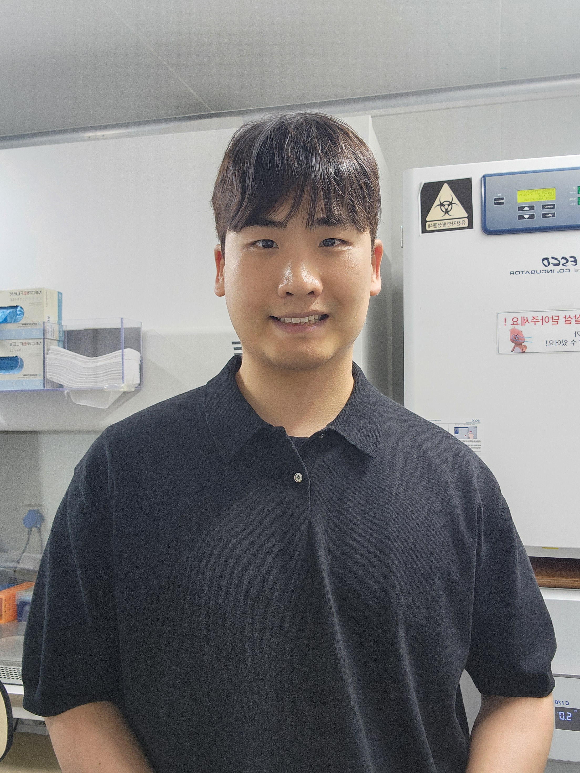
The immune system has the capability to destroy cancer cells harboring mutated genes. Cells display peptides derived from these mutated genes (i.e., portions of the mutant protein) on a molecule called the major histocompatibility complex I (MHC I), triggering cytotoxic T cells to eliminate the cancer cells. Unfortunately, this surveillance system is weak and often subverted by cancer cells. Dr. Lee [Suzanne and Bob Wright Fellow] aims to enhance the immunogenicity of the MHC I-displayed peptides using haptens, small molecules that elicit an immune response when attached to a larger carrier protein. By empowering the immune system, he envisions that these hapten-protein complexes will enable the repurposing of cancer drugs for which resistance has emerged. Dr. Lee received his PhD and BS from the Ulsan National Institute of Science and Technology, Ulsan.
Michael V. Gormally, MD, PhD
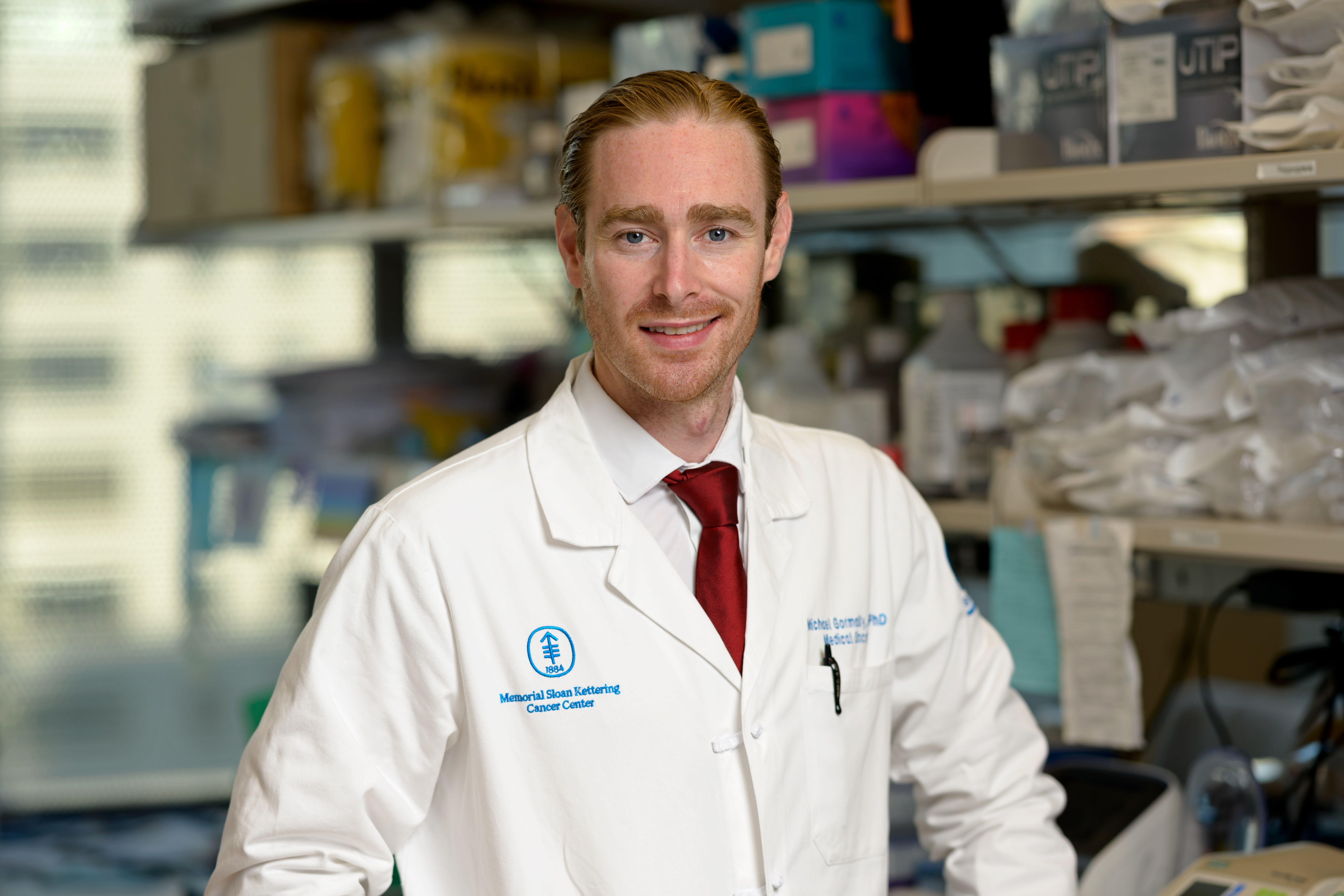
Adoptive cell therapy (ACT) is poised to expand the curative potential of immunotherapy. ACT works by administering T cells that have been genetically engineered to express tumor-specific T cell receptors (TCRs) so that they recognize a particular cancer antigen. Dr. Gormally’s [Dennis and Marsha Dammerman Fellow] work addresses two major challenges that currently limit the effectiveness of ACTs against solid tumors: identifying antigen targets that can be recognized by the immune system, and designing TCRs that target those antigens with exquisite specificity. Dr. Gormally and his colleagues have identified multiple immunogenic antigens derived from cancer-causing mutations and developed a powerful approach to retrieve potent, antigen-specific TCRs from large libraries of blood samples from cancer patients. The goal of these efforts is to identify safe and effective TCRs for clinical application. Dr. Gormally received his PhD from the University of Cambridge, Cambridge, his MD from Yale School of Medicine, New Haven, and his BA from Pomona College, Claremont.
R. Camille Brewer, PhD
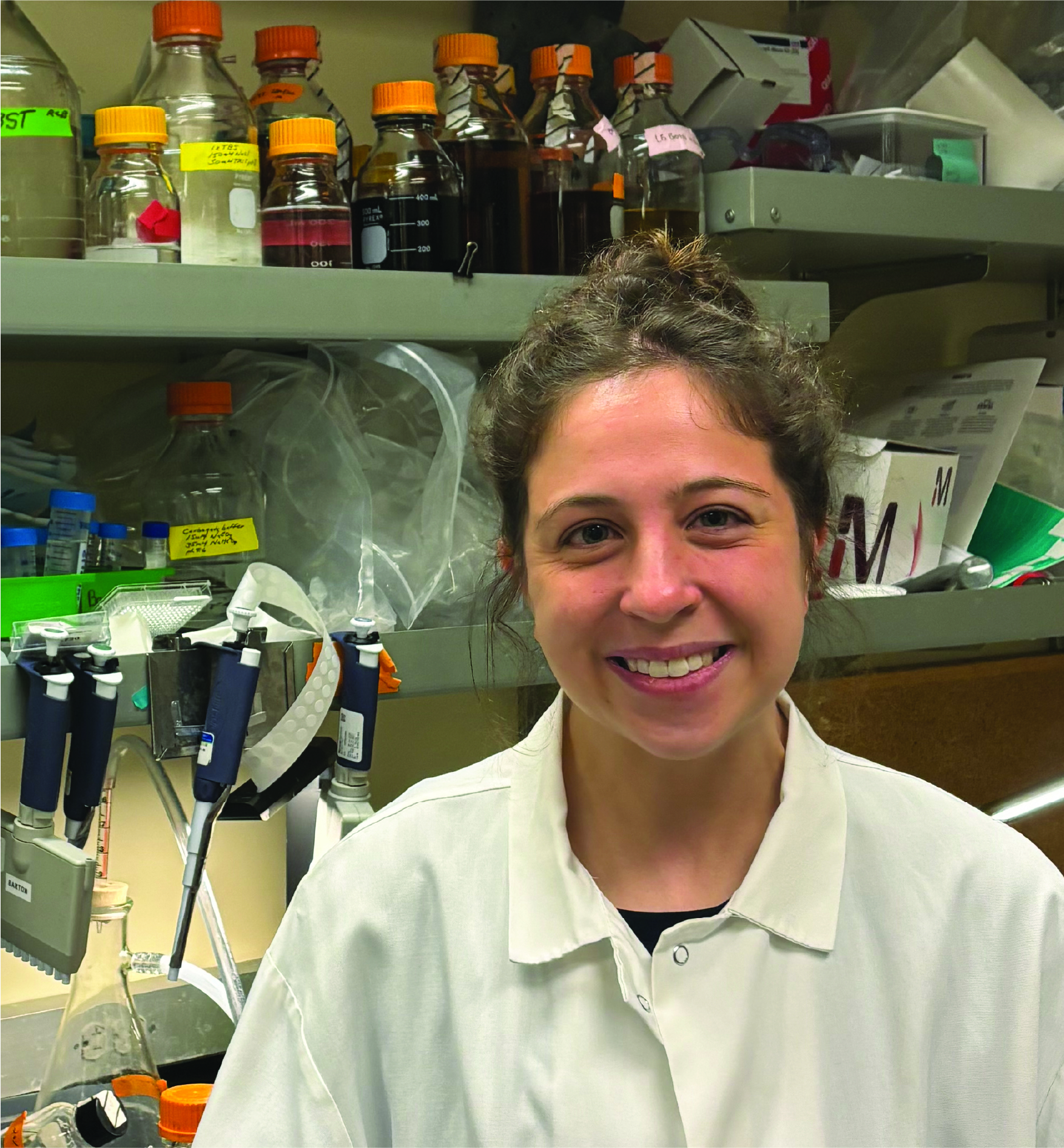
B cells, especially those that target cancer antigens, are crucial for fighting tumors; however, not everyone develops them. Our gut bacteria play a vital role in training B cells to recognize a wider range of threats. Dr. Brewer’s [HHMI Fellow] research explores how these gut bacteria influence the specificity of B cells, and thus our body’s ability to combat tumors. Dr. Brewer’s research aims to determine if the “training” of B cells by gut bacteria early in life influences their later responses to vaccines and cancer. This investigation may not only improve our understanding of how gut bacteria shape our immune system, but also pave the way for novel cancer treatments utilizing gut bacteria. Dr. Brewer received her PhD from Stanford University, Stanford and her BS from Massachusetts Institute of Technology, Cambridge.
