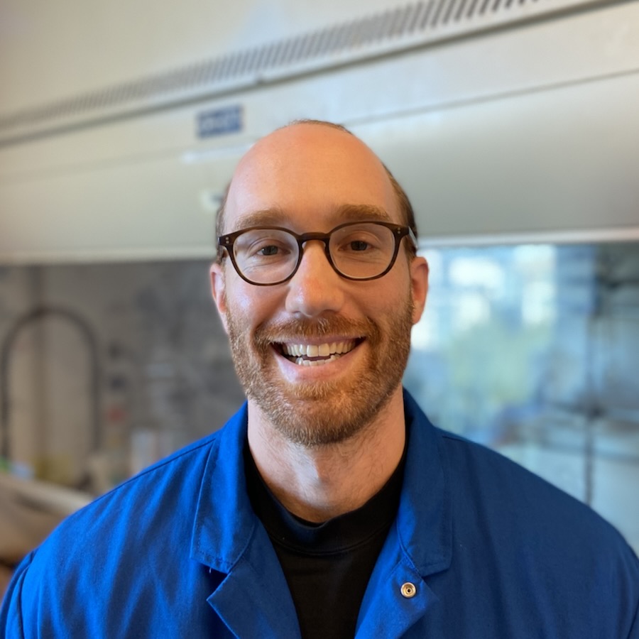
Imani Williams

Imani was born and raised in Newport News, Virginia. She attended Howard University, Washington, D.C., as a first-generation college student, earning her BS in Biology with Chemistry and Psychology minors. After graduation, she was selected as a trainee for the New York Research and Mentoring for Post Baccalaureates (NY-RaMP) Program at Hunter College, New York. She worked in Dr. Jill Bargonetti’s (Damon Runyon Fellow ‘91-’94) lab, where she focused her efforts on the study of triple-negative breast cancer research and became passionate about cancer research. She is driven by the challenge of questioning conventional wisdom and exploring seemingly implausible possibilities. She is inspired by the opportunity to reshape the future of medicine, leveraging these innovations for the benefit of all patients. Imani is committed to advocating for those in need, and she believes that pursuing cancer research will allow her to make the most meaningful impact in society.
Rebecca Pasquarelli, PhD

In order to survive, cancer cells must evade or disable the immune system. Many cancers are caused by chronic infection with viruses that modulate the host cell by influencing gene expression to achieve this same goal. Dr. Pasquarelli [HHMI Fellow] is studying how viral proteins alter gene expression to create a tumor microenvironment that prevents the immune system from killing cancer cells. Since many viral proteins achieve this by changing the expression of secreted proteins, which can be therapeutically targeted, Dr. Pasquarelli is also exploring the effect of secreted proteins on tumor immune evasion. This work will deepen our understanding of how cancers evade the immune system and has the potential to uncover new targets for cancer treatment. Dr. Pasquarelli received her PhD from the University of California, Los Angeles, and her BS from Brown University, Providence.
Anna Karen Orta, PhD

Mitochondria are best known as the cell’s power plants, but they also help cells respond to stress and repair their own DNA. A mitochondrial protein called ATAD3A plays a key role in these processes and is found at abnormally high levels in many aggressive cancers such as glioblastoma, breast cancer, and colorectal cancer, where it contributes to tumor growth and resistance to treatment. Dr. Orta studies how ATAD3A acts as a sensor of mitochondrial DNA damage—detecting trouble inside the mitochondria and helping signal to other parts of the cell, like the endoplasmic reticulum, that stress responses are needed. Using cryo-electron microscopy along with biochemical and cell-based approaches, she aims to uncover how ATAD3A is regulated and how its function supports cancer cell survival. Ultimately, she hopes to expose new ways to target mitochondrial stress pathways in cancer. Dr. Orta received her PhD from California Institute of Technology, Pasadena, and her BS from the University of Texas at El Paso, El Paso.
Léa Montégut, PhD

Early detection of lung cancer is associated with significantly better clinical outcomes. For this reason, CT-scan-based screenings in at-risk populations have been widely adopted, notably in people over 50 with a history of smoking. Still, other contributing factors include chronic exposure to environmental carcinogens, advanced age, and preexisting lung conditions. Compounding this complexity, not all precancerous lesions will evolve into invasive tumors, emphasizing the need to understand the mechanisms that govern the shift from benign to malignant states. To address this gap, Dr. Montégut [National Mah Jongg League Fellow] will focus on decoding the early immune system alterations that occur within the lung microenvironment during the pre-cancer-to-cancer transition. By doing so, she aims to identify molecular markers that indicate high-risk patients and pinpoint potential molecular targets, with the goal of intercepting tumors at a non-invasive stage. Dr. Montégut received her PhD from Paris-Saclay University, Paris, her MS from Polytechnique Montréal, Montréal, and her MEng from Ecole Polytechnique, Palaiseau.
Phaedra C. Ghazi, PhD

An emerging hallmark of cancer is phenotypic plasticity, which enables cancer cells to change their traits throughout tumorigenesis and in response to targeted therapy treatment. While new targeted therapies are emerging for the treatment of lung cancers, it has been demonstrated that lung tumors with heterogeneous cell populations are especially resistant to current treatments. One reason that lung cancer cells with the same cancer-causing mutation but different cellular identities may have differential sensitivities to targeted therapies is altered gene expression. Dr. Ghazi aims to characterize how these hybrid lung tumors are innately resistant to treatment by determining differences in gene expression and regulation. She is engineering a novel mouse model that reports the cellular identity of lung tumors in live animals. Together these efforts aim to improve our ability to treat particularly aggressive lung tumors. Dr. Ghazi received her PhD and MS from the University of Utah, Salt Lake City, and her BS from the University of Massachusetts, Amherst.
Shreoshi Sengupta, PhD

Studies have shown that lung tumors are sustained through the formation of new blood vessels from pre-existing ones in a process called angiogenesis. Moreover, tumor cells secrete signaling proteins that help them communicate with each other and evade immune detection. However, most of these studies have been on late-stage lung tumors; our understanding of cell-cell interactions in the tumor environment during lung cancer initiation and early stages remains poor. Dr. Sengupta [Deborah J. Coleman Fellow] plans to identify the gene expression patterns in tumor cells, endothelial cells (blood-vessel-forming cells), and immune cells over time to understand how they engage in this cellular crosstalk, promoting tumorigenesis. She also plans to examine cell-cell interactions in early-stage lung cancer using organoids, or artificially grown miniature organs. This line of investigation will help understand the mechanisms underlying tumor initiation and lead to novel biomarkers that can help detect lung cancers earlier. The findings will also help identify novel therapeutic targets that can be inhibited to improve patient responses and survival. Dr. Sengupta received her PhD from Indian Institute of Science, Bangalore and her MS and BS from University of Calcutta, Kolkata.
Saket Rahul Bagde, PhD

Most cancers develop in the epithelial tissue, which includes the skin and internal organ linings. Hemidesmosomes (HDs) are adhesive structures that anchor epithelial cells to the underlying base layer and maintain tissue integrity. While HD disassembly occurs normally during wound healing, tumor cells can exploit this process to detach and spread to other parts of the body. Dr. Bagde [Bakewell Foundation Fellow] is studying how HD components interlock like Lego blocks to form stable HDs in healthy tissues and how they disassemble in cancerous tissues. To investigate this phenomenon, Dr. Bagde plans to develop organoids—self-organizing mini-organs grown in a petri dish to study disease progression. By creating simple base layers that simulate the supportive properties of the native organ base layer, he plans to promote the growth of both normal and cancerous organoids. This work has the potential to support the development of personalized cancer therapies based on patient-derived tumor samples. Dr. Bagde received his PhD from Cornell University, Ithaca and his MS and BS from the Indian Institute of Science Education and Research, Pune.
Rodrigo Gier, PhD

Drug therapies that selectively target proteins that drive the growth of tumor cells are rapidly becoming the standard of care for many cancers. However, tumors are often able to evade inhibition by targeted anti-cancer drugs by activating other proteins, leading to drug resistance. Dr. Gier [HHMI Fellow] is developing a new therapeutic approach that repurposes existing drugs to release highly toxic cargoes, known as payloads, that aggregate in drug-resistant cancer cells and kill them. As a general platform, it is applicable to a wide range of solid and liquid cancers. Dr. Gier received his PhD from University of Pennsylvania, Philadelphia and his BA from Swarthmore College, Swarthmore.
Laura Crowley, PhD

Fibroblasts are one of the earliest known cell types and they contribute to many of the most burdensome lung diseases, including cancers, fibrosis, and emphysema; however, they are surprisingly poorly understood. Dr. Crowley [HHMI Fellow] will examine the different types of fibroblasts in the mouse lung to determine where they come from and how they function normally, as well as how they change with injury and disease. This will establish an important baseline for how these cells function in mice and also provide critical, long-term insights into how these cells may function in humans, where lung diseases are very difficult to treat and are among the leading causes of mortality worldwide. Though her work will directly analyze the fibroblasts and microenvironment around lung tumors, her findings could translate to many other solid tumor contexts. Dr. Crowley received her PhD from Columbia University, New York and her BA from Colby College, Waterville.
Pu Zheng, PhD

Dr. Zheng [Fayez Sarofim Fellow] is dedicated to the development of technologies for studying tumor evolution within their native contexts. Understanding the complex processes of cancer growth and progression requires a deep exploration of the dynamic interactions between tumor cells and the tumor microenvironment. “Spatial-omics” technologies are powerful tools that offer direct visualization of cells and their interactions in natural contexts, enabling systematic investigation of these intricate processes. Dr. Zheng aims to develop novel spatial-omics technologies that combine imaging and gene sequencing approaches to uncover the mechanisms underlying the spatially distinguished features of tumor evolution. Dr. Zheng received his PhD from Harvard University, Cambridge and his BS from Peking University, Beijing.
