The rise of single-cell RNA sequencing in recent years has transformed the study of gene expression, providing researchers with a detailed picture of how and when genes get turned “on” and “off” in individual cells within a given tissue. Analyzing cells’ RNA sequences, or transcriptomes, can reveal cell-to-cell variability, or in the case of cancer, mutations carried by small populations of tumor cells. Current single-cell sequencing methods, however, fail to capture the location of the cell within the tissue. Spatial transcriptomics techniques, on the other hand, define the spatial distribution of RNA molecules within a tissue sample, but lack single-cell resolution. To put this on a human scale, consider the different information you get about a neighborhood from a phone book versus a satellite image.

At MD Anderson Cancer Center, Damon Runyon Quantitative Biology Fellow Runmin Wei, PhD, and former Damon Runyon Innovator Nicholas E. Navin, PhD, have developed the first tool that combines these two datasets, creating spatial maps of tissue samples at single-cell resolution. The tool, known as CellTrek, offers detailed information about individual cells’ behavior from RNA-sequencing data while showing their location and microenvironment within the tissue. This approach allows researchers to gauge the degree of heterogeneity within a tumor as well as pinpoint possible targets within specific regions.
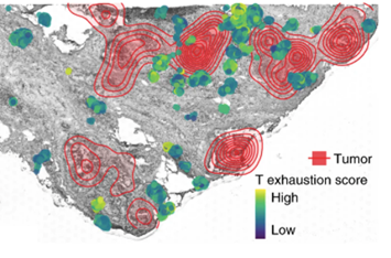
By applying CellTrek to samples of breast cancer tissue, for example, the team noticed that immune cells clustered unevenly around different subgroups of tumor cells. Some tumor cells had more “exhausted” T cells near them than others, suggesting that some genetic or molecular feature of those cells has an immunosuppressive effect. These cells might be prioritized as targets in order to slow tumor growth.
“While this approach is not restricted to analyzing tumor tissues, there are obvious applications for better understanding cancer,” says Dr. Navin. “Pathology really drives cancer diagnoses and, with this tool, we’re able to map molecular data on top of pathological data to allow even deeper classifications of tumors and to better guide treatment approaches.”
This research was published in Nature Biotechnology.


